Chronic Ear Infections and Total Ear Canal Ablation

Chronic ear infections are a common and frustrating disease for owners and veterinarians. For the pet, however, the condition is much more than frustrating. Pets suffering from chronic ear infections and inflammation are often in severe pain, and both cats and dogs can be affected. Chronic ear infections would be defined as multiple infections per year or infections that never really resolve.
Also referred to as chronic otitis, this condition affects some breeds more frequently than others. Cocker Spaniels are predisposed, as are other spaniel breeds, Labrador Retrievers, and other breeds with “floppy” ears. This is probably due both to the conformation of the ear as well as tendency for those breeds to have allergies – the perfect situation for development of chronic ear infections.
Signs of otitis depend on the severity of the infection and degree of inflammation but may include:
- Shaking the head
- Scratching at the ears
- Rubbing the head and ears on the floor or furniture
- Crying or groaning as they rub and scratch ears
- Discharge from the ears, which can sometimes have a foul odor
- Redness of the ear canal and ear flap (the ears may also feel warm when touched)
- Ear hematoma, evidenced by a severely swollen ear flap
- Aggression or avoidance when the head or ears are approached
Appropriate medical management is often successful in curing acute otitis. Unfortunately, treatment often only works temporarily. Painful ears can prove challenging to medicate, and if the underlying cause of the infection is not addressed, the treatment may fail to work at all. Once the condition becomes long-term, or “chronic”, treatment becomes increasingly difficult to manage and antibiotic resistance also becomes a concern.
The underlying cause of otitis is often allergic. These patients get stuck in a cycle of inflammation and infection which eventually leads to thickening and scarring of the tissues, collapse of the ear canals, ruptured ear drums, and accumulation of debris and infection within the middle ear. Over time, the scar tissue blocks the canals, preventing topical medications from reaching the diseased portions. The blocked canals also prevent the natural shedding of wax and hair. This debris accumulates in the canals and within the middle ear, adding to inflammation and pain.
We see many of these cases here at VMC, where the cycle of pain, itching, chronic pus and infection, head shaking, chronic medication, and owner fatigue have led the pet owner to seek a surgical specialist and a permanent solution.
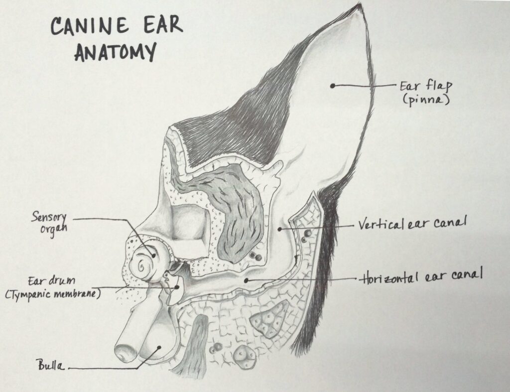
A Total Ear Canal Ablation (or TECA) is a procedure that removes the vertical and horizontal ear canals down to the level of the middle ear. Due to the high incidence of middle ear involvement with chronic otitis, the middle ear is also cleaned out using a technique called a lateral bulla osteotomy. That procedure removes some bone and any infected tissue to allow for continued healing. While TECA procedures are most often performed to treat severe cases of chronic otitis and infection, the procedure is also performed in the treatment of other ear diseases, such as traumatic ear canal injury and when removing tumors in the ear.
This may sound dramatic, but the permanent treatment is much better than the chronic pus, pain, and constant medicating that most pets are experiencing. During the surgery, it is normal to find a large amount of debris, hair, and pus in the bulla. It is easy to understand then, why these cases of chronic disease have not resolved medically, given the amount of debris within the middle ear. We submit samples of the tissues in the bulla to a lab for culturing, and we commonly find that antibiotic resistant bacteria (such as MRSA) are present.
The most common complications with the TECA are facial nerve paralysis and vestibular problems or “vertigo.” Facial nerve paralysis and vertigo are usually temporary and resolve without specific treatment. A very small percentage of cases can experience an abscess in the area months or years later. During surgery, the bulla cavity is cleaned of all recognizable tissues, but if any microscopic cells remain, they have the potential to grow and begin to shed cells again. This debris can accumulate in the bulla, sometimes becomes infected, and may develop an abscess. All associated risks increase with the severity of the condition, so considering this treatment early in the disease process is recommended.
As with any other surgery, there is a risk of anesthetic complication. In general, most anesthetic problems can be prevented with complete assessment of the patient. The physical examination and preoperative bloodwork will provide baseline information and guide recommendations for additional diagnostic testing prior to anesthesia. All surgical patients at VMC are monitored using multi-parameter equipment and a Licensed Veterinary Technician dedicated to your pet. There are no anesthesia complications unique to TECA surgery, although each patient is unique with his/her own limitations and concerns to be managed.
Many owners are concerned about deafness after the surgery. While the TECA removes the apparatus that transmits sound via the air (i.e., the ear canal and ear drum) sound can still be sensed via the vibrations that come to the cochlear apparatus through the sinuses and skull. This is similar to the level of hearing one experiences when wearing earplugs or when underwater. No sound reaches the cochlear apparatus through the air, but we can still hear sounds and voices. The reality is that most dogs with chronic otitis are already hearing at this low level due to the collapse and obstruction of their ear canal and middle ear, where no sound waves are being transmitted via the air. Most owners do not report a change in the pet’s ability to hear after a TECA.
A TECA surgery is often a very rewarding surgery for the patient, the pet owner, and the veterinarian. Most owners report a dramatic improvement in the attitude of their pets post-operatively, and they frequently report an increase in activity level and play behaviors – sometimes behaviors they have not seen in months or years. A more social and playful pet, combined with relief from daily ear cleaning and medicating, offers the owner a huge sense of relief.

In summary, a TECA surgery will:
- Eliminate ongoing ear pain, odor, and infection
- Eliminate the need for and cost of ear medications and veterinary visits to treat chronic ear infections
- Improve your relationship with your pet (no odor, no pain, no difficult ear treatments)
- Improve your pet’s attitude, activity and general well being with the removal of chronic pain, inflammation, and infection
Some pets, particularly those with long ear flaps, may still require topical cleaning post-surgically. Surgery will not correct the allergic reaction that creates a good environment for bacteria and yeast to grow under those flaps (or ear pinnae), but, overall, the pet’s comfort will be much increased.
The TECA should not be viewed as strictly a salvage procedure or last resort. The benefits of the procedure argue for it to be recommended much earlier in the course of the disease process. Many dogs and cats with chronic otitis are excellent candidates for the surgery once it becomes clear that the otitis is a chronic problem. Additionally, potential complications will be fewest if the surgery is performed early in the disease process, and both you and your pet can appreciate the benefits earlier!
READ THIS POST FOR THE STORY OF A DOG WHO HAS BENEFITED FROM THIS SURGERY!
WHAT HAPPENS DURING SURGERY?
After your pet is under anesthesia, his/her ears flaps and surrounding skin are shaved, and the ear canal is cleaned. The area is prepared for sterile surgery.
An incision is made around the cartilage of the external/outer ear canal, and the canal is removed down to the level of the middle ear. The middle ear (also called the “bulla”) is a boney, eggshell-like structure on the side of the skull. The eardrum normally is stretched across the opening between the external/outer ear and the middle ear. In the majority of dogs requiring a TECA, the ear drum has been ruptured for a long period of time. The middle ear is cleaned of infected material and tissue, and then flushed with sterile saline. A culture sample is taken at this time to help identify any remaining bacteria.
The surgery is completed by sewing the tissues closed in several layers, ending with sutures in the skin completely closing what used to be an open ear canal. A temporary drain will be left in place for 1-2 days.
WHAT HAPPENS AFTER SURGERY?
Your pet will stay in the hospital for at least 24hrs so we can manage recovery and pain control. Patients are often more comfortable if their head and ears are bandaged snuggly to prevent ear flapping or bumping the incision.
An antibiotic will be prescribed for up to 2-4wks. Pain medications will also be prescribed for the first week post-operatively.
Sutures will need to be removed 10-14 days after surgery.
READ THIS POST FOR THE STORY OF A DOG WHO HAS BENEFITED FROM THIS SURGERY!
About Us
At Veterinary Medical Center of Central New York, we understand that the health of your pet is a top priority. In the case of an emergency or a condition requiring specialized care, we're here to treat your pet in our state-of-the-art, 24-hour Emergency and Critical Care Center.